20 Dec The Role of Brain Imaging Techniques (MRI And FMRI) in ADHD Research
INSIDE THE ADHD BRAIN:
What Advanced Imaging Reveals

Ever wonder what’s going on inside the brain of someone with ADHD? Thanks to recent advances in brain imaging techniques like MRI and fMRI, scientists are getting an unprecedented view into how the ADHD brain works—and doesn’t work. If you or someone you know lives with ADHD, this glimpse into the inner workings of the ADHD brain may help explain some of the daily struggles and frustrations. But it also offers hope. We get closer to more targeted treatments as we better understand the underlying issues.
The Role of Brain Imaging Techniques MRI and fMRI in ADHD Research
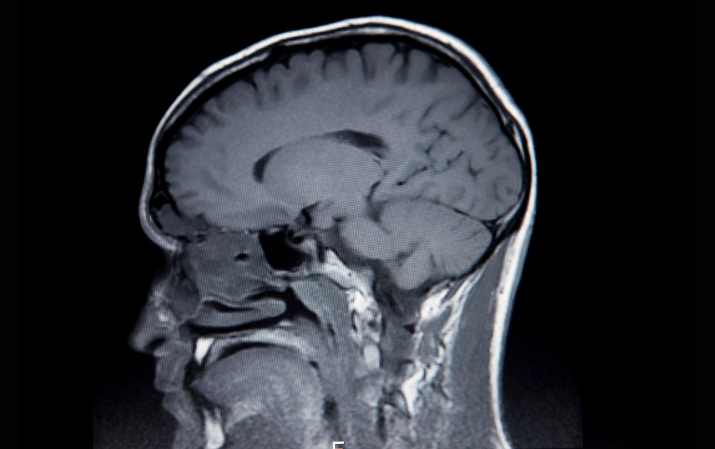
Functional MRI (fMRI) and structural MRI have allowed scientists to peer inside the ADHD brain like never before. fMRI, in particular, can detect changes in blood flow as certain brain areas activate, giving researchers insights into how the ADHD brain functions differently.
Studies using fMRI show that in individuals with ADHD, the areas of the brain involved in focus and attention, like the prefrontal cortex, tend to be less active. At the same time, areas involved in reward and motivation, like the ventral striatum, appear hyperactive. This combination makes it hard to focus but easy to get distracted by exciting or interesting things.
fMRI also reveals that the ADHD brain has a harder time connecting the prefrontal cortex and ventral striatum. The prefrontal cortex helps control impulses and make good decisions, while the ventral striatum is involved in motivation and reward. A weaker connection between these areas may make it harder for someone with ADHD to self-regulate.
Other studies show that the ADHD brain has lower dopamine levels, an important neurotransmitter for motivation and focus. Medications for ADHD, like stimulants, work by increasing dopamine levels in the brain to improve functioning in critical areas.
While fMRI and MRI have revealed biological differences that contribute to symptoms of ADHD, the diagnosis still relies primarily on behavioral assessments. However, as technology and understanding advance, brain scans could play an increasing role in diagnosis and treatment. A more personalized approach may help tailor treatments to individual needs based on their unique brain function.
The future is bright for gaining a better understanding of ADHD’s neurological underpinnings. Continued research into the structural and functional variations of the ADHD brain can pave the way for earlier diagnosis, more targeted interventions and new treatment options to help those affected live their best lives.
Structural Differences in the ADHD Brain Revealed by MRI
When researchers peer into the ADHD brain using MRI and fMRI scans, they uncover some striking differences in both structure and function compared to neurotypical brains.
MRI scans reveal that several areas of the ADHD brain, including the prefrontal cortex, cerebellum, and basal ganglia, tend to be smaller in size and volume. The prefrontal cortex, responsible for executive functions like planning and impulse control, is particularly impacted. This likely contributes to difficulties with focus, organization, and decision-making that many with ADHD experience.
fMRI scans go a step further by measuring brain activity and blood flow. These functional scans show that the ADHD brain also has lower activity in the prefrontal cortex and other frontal lobe regions involved in self-regulation and reward processing. At the same time, some deeper brain regions involved in motivation and reward show higher activity. This combination may drive tendencies toward impulsiveness, hyperactivity, and seeking stimulation or rewards.
Neurotransmitters, the chemical messengers in the brain, are also implicated in ADHD. Studies point to imbalances or deficiencies in dopamine and norepinephrine, neurotransmitters important for attention, motivation, and reward processing. Stimulant medications for ADHD work by increasing dopamine and norepinephrine activity in the prefrontal cortex and connected regions to improve functioning.
While more research is needed, advanced imaging and an evolving understanding of the neurochemistry behind ADHD are helping to improve diagnosis and open doors to new treatment options. The future is bright for better understanding and managing this disorder.

Functional Variations in the ADHD Brain Uncovered by fMRI
Functional MRI (fMRI) scans allow researchers to see the ADHD brain in action. These scans measure changes in blood flow in the brain, which indicates which areas are more or less active during certain tasks. Studies using fMRI have found some significant differences in how the ADHD brain functions.
One of the hallmark signs of ADHD is difficulty with executive function, the ability to plan, organize, and regulate behavior. fMRI scans show less activity in the frontal lobe of the ADHD brain during tasks involving executive function, like decision-making or impulse control. There also seems to be a lack of connectivity between the frontal lobe and other areas involved in executive function, like the basal ganglia.
fMRI scans also reveal decreased activity in the basal ganglia, which is important for motivation and reward processing. This may explain why people with ADHD have trouble staying focused on tasks that aren’t highly stimulating or rewarding. The basal ganglia interact closely with the dopamine system, and dopamine is thought to play an important role in ADHD.
Other studies have found decreased activity in the anterior cingulate cortex and caudate nucleus, areas involved in emotional regulation and motivation. There also seems to be a lack of connectivity between these areas and the prefrontal cortex. These findings provide biological evidence for the inspirational and motivational symptoms of ADHD.
In summary, fMRI has been invaluable for identifying functional differences that provide insight into the underlying mechanisms of ADHD. Continued research using fMRI and other imaging techniques will help improve diagnosis and open doors to new treatment options aimed at targeting the specific areas of deficiency in the ADHD brain.
The Role of Neurotransmitters in ADHD
When it comes to ADHD, neurotransmitters—the chemical messengers in your brain—play an important role. Two key neurotransmitters involved in ADHD are dopamine and norepinephrine.
Dopamine
Dopamine is crucial for motivation, reward-seeking behavior, and motor control. For people with ADHD, dopamine signaling seems to be impaired, making it difficult to focus and control impulses. Imaging scans show differences in the dopamine system of the ADHD brain compared to neurotypical brains.
Medications for ADHD, like stimulants, work by increasing dopamine levels in the brain. This helps improve focus and attention while reducing hyperactive and impulsive behaviors.
Norepinephrine
Norepinephrine is involved in arousal, alertness, and stress responses. It seems norepinephrine signaling may also be altered in ADHD, contributing to problems with attention regulation and impulsiveness.
Some ADHD medications also target the norepinephrine system. Reuptake inhibitors for norepinephrine and dopamine, known as SNRIs, can help improve focus and concentration in people with ADHD.
While neurotransmitters are not the only factor involved in ADHD, they do appear to play a significant role for many individuals. A better understanding of the neurochemistry behind ADHD is helping to guide treatment and may eventually improve diagnosis.
The more we understand the inner workings of the ADHD brain, the better equipped we are to develop targeted interventions. Advanced imaging and an expanding knowledge of neurochemistry continue to provide insight into this complex condition.

Implications for ADHD Treatment and Management
The discoveries from advanced brain imaging have significant implications for how we understand and treat ADHD. Knowledge about structural and functional differences can help guide more targeted treatment approaches.
Medication
Medications for ADHD work by altering neurotransmitter levels, especially dopamine and norepinephrine, to improve focus and self-control. Imaging shows these meds can normalize activity in the prefrontal cortex and striatum, areas involved in executive function and reward processing. Understanding a patient’s specific brain differences may help determine which meds and dosages could be most effective for them.
Behavioral Interventions
Non-drug interventions like cognitive behavioral therapy (CBT) can also benefit from neuroimaging insights. CBT aims to modify problematic thoughts and behaviors. Knowing which brain regions are most impacted in a patient allows therapists to tailor strategies to target executive functions like planning, prioritizing, and impulse control specifically. Mindfulness practices also help by increasing awareness and control over wandering attention.
Early Intervention
Identifying ADHD-related brain changes can allow for earlier diagnosis and treatment. The brain is most malleable in children, so intervening early with therapy and meds may help rewire connections before symptoms worsen. Studies show treatment can significantly improve the brain development trajectory.
Managing Severe Cases
For severe ADHD that does not respond sufficiently to typical treatments, alternative options may be explored, such as neurofeedback training. This technique provides real-time feedback about brain wave activity to help people learn to regulate their brain function better. While still considered experimental, it shows promise for normalizing the brain activity patterns associated with ADHD symptoms.
The future is bright for better understanding and managing ADHD. Continued research on brain structure and function will unlock new insights into this complex disorder and open more doors to personalized, effective solutions. The key is to keep exploring, learning, and applying the latest knowledge to help improve lives.
Conclusion
So there you have it, an inside look at how ADHD impacts the structure and function of your brain. While ADHD is often thought of as a behavioral issue, we now know it has real neurological underpinnings. The good news is we have technologies like MRI and fMRI to visualize what’s happening in the ADHD brain. These scans are helping researchers better understand the role of neurotransmitters like dopamine and norepinephrine. They’re also shedding light on differences in connectivity and activity patterns in key focus, attention, and inhibition regions. The more we learn, the better equipped we’ll be to diagnose and treat the condition. The future is bright as we work to unlock the mysteries behind that restless, distracted mind of yours. Keep your head up – the answers are coming into focus.
Craig Selinger
Latest posts by Craig Selinger (see all)
- Psychotherapy and Support Services at Cope With School NYC - April 12, 2024
- NYC Parents of Teens Support Group - April 8, 2024
- Here I Am, I Am Me: An Illustrated Guide to Mental Health - April 4, 2024


No Comments